Beskrivelse


FUNDUS CAMERAType Non-mydriatic fundus camera
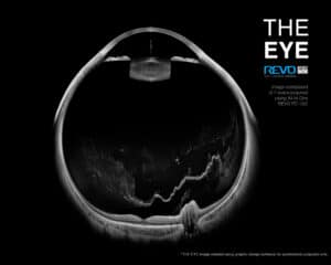
Photography type Color
Angle of view 45° ± 5% or less
Min. pupil size for fundus 3.3 mm or more
Camera 12.3 Megapixel CCD camera
Camera modes PhotographyFundus (Retina, Central, Disc, Manual fixation), Anterior photo
Flash adjustment, Gain, Exposure Auto, Manual
Intensity levels High, Normal, Low
OPTICAL COHERENCE TOMOGRAPHY:
Spectral Domain OCT
Light Source SLED, Wavelength 850 nm
Bandwidth 50 nm half bandwidth
Scanning speed 80 000 measurements per second
Axial resolution 2.6 μm digital, 5 μm in tissue
Transverse Resolution 12 μm, typical 18 μm
Overall scan depth 2.8 mm / ~6 mm in Full Range mode
Min. pupil size for OCT 2.4 mm
Focus adjustment range -25 D to +25 D
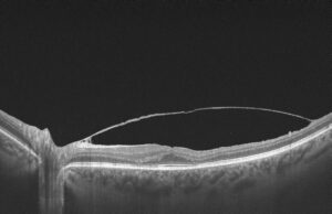
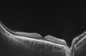
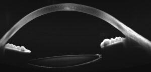
Scan range Posterior 5 mm to 15 mm, Angio 3 mm to 12 mm, Anterior 3 mm to 18 mm
Scan types 3D, Angio*, Full Range Radial, Full Range B-scan, Radial (HD), B-scan (HD), Raster (HD), Cross (HD), TOPO, AL, ACD
Fundus alignment Live Fundus Reconstruction
Alignment method Fully automatic, Automatic, Manual
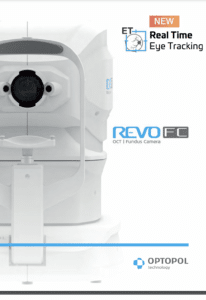
Fundus Tracking real time , iTracking
Retina analysis Retina thickness, Inner Retinal thickness, Outer Retinal thickness, RNFL+GCL+IPL thickness, GCL+IPL thickness, RNFL thickness, RPE deformation, MZ/EZ-RPE thickness
Angiography OCT
an optional software module to purchase Vitreous, Retina, Choroid, Superfi cial Plexus, RPCP, Deep Plexus, Outer Retina, Choriocapilaries, Depth Coded, SVC, DVC, ICP, DCP, Custom, Enface, FAZ, VFA, NFA, Quantifi cation: Vessel Area Density, Skeleton Area Density, Thickness map
Glaucoma analysis RNFL, ONH morphology, DDLS, OU and Hemisphere asymmetry, Ganglion analysis as RNFL+GCL+IP and GCL+IPL,
Structure + Function
Angiography mosaic Acquistion method: Auto, Manual
Mosaic modes: 10 x 6 mm, Manual up to 12 images
Biometry OCT
an optional software module to purchaseAL, CCT, ACD, LT, P, WTW
IOL Formulas: Hoffer Q, Holladay I, Haigis, Theoretical T, Regression II
Corneal Topography Map
an optional software module to purchaseAxial [Anterior, Posterior], Refractive Power [Kerato, Anterior, Posterior, Total], Net Map, Axial True Net, Equivalent Keratometer, Elevation [Anterior, Posterior], Height, KPI (Keratoconus Prediction Index)
Anterior
No adapter required even for wide scans e.g. Angle to AngleAnterior Chamber Radial, Anterior Chamber B-scan, Pachymetry, Epithelium map, Stroma map, Angle Assessment, AIOP, AOD 500/750, TISA 500/750, Angle to Angle view, Wide Angle, Wide Cornea
Connectivity DICOM Storage SCU, DICOM MWL SCU, CMDL, Networking
Fixation target OLED display (The target shape and position can be changed), External fixation arm
Dimensions (LxWxH) / Weight 479 × 367 × 493 mm / 30 kg
Power supply / consumption100-240 V, 50/60 Hz / 90-110 VA